.svg) National Institute of General Medical Sciences |
 |
|
National Biomedical Resource for |
| ACERT's Service and Collaborative Projects | |
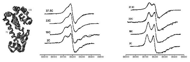
Studies of structure and dynamics of proteins are important for the understanding of protein function. Site directed spin labeling (SDSL) has proven to be useful for such studies. However, conventional ESR studies of protein dynamics are limited by the resolution of the spectrum to the molecular dynamics. This is especially true when one considers the complexity of protein dynamics. That is, the rotational dynamics of a nitroxide spin tethered to the protein at the labeled site includes several types of motions: 1) internal motion of the nitroxide, 2) backbone fluctuations, and 3) global rotation of the protein. The challenge is in unraveling these types of motions and describing them with accurate motional and ordering parameters. We have developed a multi-dimensional ESR approach, wherein high frequency (250 GHz) ESR is used as a "fast snapshot" of the dynamics, wherein the slow global rotational diffusion of the protein appears to be frozen out, but the spectral lineshape is particularly sensitive to the faster internal modes of motion. On the other hand, a low frequency (9 GHz), or conventional, ESR spectrum corresponds to a "slower snapshot" of the dynamics, so it is also sensitive to the global diffusion. This approach has been utilized to study T4 Lysozyme spin labeled at different sites. A particularly relevant comparison is afforded by labels at sites 131 and 72 shown in the Figure below on the left. Their respective 250 GHz ESR spectra are shown below (131 on the left and 72 on the right). These spectra are distinctly different. Since the 250 GHz spectra are sensitive to just the internal modes of motion, it is clear that these sites must have different structural dynamic characteristics. The respective 9 GHz spectra, which are affected by the same global motion of the protein, do not display such clear spectral differences. The spectra at the different frequencies have been simultaneously fit to the slowly relaxing structure model, which includes these various types of motions, and these fits are shown below for the 250 GHz spectra. We have found that the key difference between these two sites is that the 72 site is more highly ordered than the 131 site, whereas their local motional rates are similar. This is consistent with the fact that whereas at both sites the side chain containing the nitroxide makes no tertiary contacts, site 72 is at the center of a long helix while site 131 is in a short two turn helix, which allows greater flexibility, i.e. less constrained motion. This study illustrates how multi-frequency ESR may be used to decompose the different modes of protein motion affecting each site as a result of their different timescales. Publication: Z. Liang, Y. Lou, J.H. Freed, L. Columbus, and W.L. Hubbell, J. Phys. Chem. B, 108, 17649-17659 (2004); no PMCID |
|
|
|
|
Z. Liang, Y. Lou, and J. H. Freed (ACERT), L. Columbus, and W. L. Hubbell (University of California, Los Angeles) |
|
|
|
About ACERT Contact Us |
Research |
Outreach |
ACERT is supported by grant 1R24GM146107 from the National Institute of General Medical Sciences (NIGMS), part of the National Institutes of Health. |
|||||
| ||||||||