.svg) National Institute of General Medical Sciences |
 |
|
National Biomedical Resource for |
| ACERT's Service and Collaborative Projects | ||
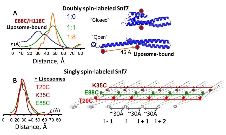
The endosomal sorting complexes required for transport (ESCRTs) constitute hetero-oligomeric machines that catalyze multiple topologically similar membrane-remodeling processes. Although ESCRT-III subunits polymerize into spirals, how individual ESCRT-III subunits are activated and assembled together into a membrane-deforming filament was unknown. Using pulsed dipolar electron spin resonance spectroscopy (PDS), we showed that Snf7 activation requires a prominent conformational rearrangement to expose protein-membrane and protein-protein interfaces. This promotes the assembly of Snf7 arrays with ~30 Å periodicity, giving rise to a membrane-sculpting filament. Then by using a combination of biochemical and genetic approaches, both in vitro and in vivo, we demonstrated that mutations on these protein interfaces abolish Snf7 assembly and block ESCRT function. The architecture of the activated and membrane-bound Snf7 polymer provides crucial insights into the spatially unique ESCRT-III-mediated membrane remodeling. This study required the use of samples with very low concentrations of labeled membrane protein, less than 10 μM, demonstrating the possibilities offered by the high sensitivity of ACERT's PDS spectrometers. Funding: P41GM105321, R01EB003150 (to JHF), T32GM007273 (to ST), R01GM094347 (to YM), R01GM098621 (to JCF); Amer. Cancer Soc. PF-12-026-01-DMC (to NJB); Cornell Univ. CU3704 (to SDE). Publication: S. Tang, W.M. Henne, P.P. Borbat, N.J. Buchkovich, J.H. Freed, Y. Mao, J.C. Fromme, and S.D. Emr. eLife 4, e12548 (2015); PMCID: PMC4720517. |
||
|
||
|
S. Tang, W.M. Henne (Weill Institute of Cell and Molecular Biology, Cornell University, Ithaca; Department of Molecular Biology and Genetics, Cornell University, Ithaca) P.P. Borbat (Department of Chemistry and Chemical Biology, Cornell University, Ithaca, NY; ACERT) N.J. Buchkovich (Weill Institute of Cell and Molecular Biology, Cornell University, Ithaca; Department of Molecular Biology and Genetics, Cornell University, Ithaca) J.H. Freed (Department of Chemistry and Chemical Biology, Cornell University, Ithaca, NY; ACERT) Y. Mao, J.C. Fromme, and S.D. Emr (Weill Institute of Cell and Molecular Biology, Cornell University, Ithaca; Department of Molecular Biology and Genetics, Cornell University, Ithaca) |
||
|
|
About ACERT Contact Us |
Research |
Outreach |
ACERT is supported by grant 1R24GM146107 from the National Institute of General Medical Sciences (NIGMS), part of the National Institutes of Health. |
|||||
| ||||||||