.svg) National Institute of General Medical Sciences |
 |
|
National Biomedical Resource for |
| ACERT's Service and Collaborative Projects | ||
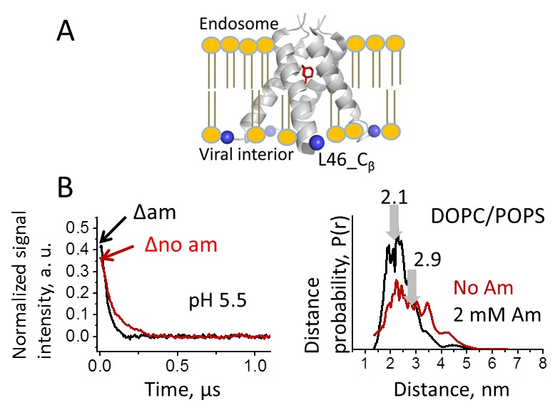
The M2 protein from influenza A plays important roles in its viral cycle. It contains a single transmembrane helix, which oligomerizes into a homotetrameric proton channel that conducts in the low-pH environment of the host-cell endosome, leading to virion uncoating at an early stage of infection. Using double electron-electron resonance (DEER) Pulse Dipolar ESR (PDS), we studied the conformational rearrangements that occur in the M2 core transmembrane domain (M2TMD) residing in the lipid bilayer, upon its interaction with amantadine drug at pH 5.5 when M2 is conductive. M2 was labeled at the residue L46C with nitroxide spin-label. First, electron microscopy using Nanogold® reagent confirmed our previous PDS results that M2 reconstituted into DOPC/POPS is an admixture of monomers, dimers, and tetramers, thus confirming our model based on a dimer intermediate in the assembly of M2. The newer PDS study showed that in DOPC/POPS membranes amantadine shifts oligomer equilibrium to favor tetramers. To do so, we used the DEER method based on accurate measurement of the modulation depth of the time-domain DEER signal, as developed in our previous study on M2, enabling the determination of the relative amounts of oligomeric species in heterogeneous protein systems residing in lipid membranes. For this characterization we used a very broad range of protein-to-lipid molar ratios (P/L's) from ca. 1:20,000 to 1:160, necessitating the use of very low protein concentrations, as low as 5 μM at the smallest P/L's. These experiments were possible only because of the very high sensitivity and stability of the PDS spectrometer at ACERT operating at 17.3 GHz. Furthermore, we found that amantadine binding shortens the inter-spin distances (for nitroxide labels) by 5-8 Å, indicating drug-induced channel closure on the C-terminal side. This effect is relevant to the mechanism of M2 inhibition. No such effect was observed for the thinner membrane of DLPC/DLPS, emphasizing the impact of bilayer thickness on protein function. Thus, the inhibited M2 channel is a stable tetramer with a closed C-terminal exit pore. Funding: P41GM105321, R01EB003150. Publication: |
||
|
||
|
E.R. Georgieva, P.P. Borbat (Department of Chemistry and Chemical Biology, Cornell University, Ithaca, NY; ACERT) H.D. Norman (School of Engineering, Cornell University, Ithaca, NY) K. Grushin, S. Stoilova-McPhie (Department of Neuroscience and Cell Biology, Sealy Center for Structural Biology and Molecular Biophysics, University of Texas Medical Branch at Galveston, Galveston, TX) N.J. Kulkarni (College of Art and Sciences, Cornell University, Ithaca, NY) Z. Liang, and J.H. Freed (Department of Chemistry and Chemical Biology, Cornell University, Ithaca, NY; ACERT) |
||
|
|
About ACERT Contact Us |
Research |
Outreach |
ACERT is supported by grant 1R24GM146107 from the National Institute of General Medical Sciences (NIGMS), part of the National Institutes of Health. |
|||||
| ||||||||