.svg) National Institute of General Medical Sciences |
 |
|
National Biomedical Resource for |
| ACERT's Service and Collaborative Projects | |
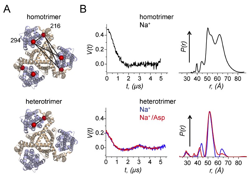
Membrane proteins such as ion channels and transporters are frequently homomeric. The homomeric nature raises important questions regarding coupling between subunits and complicates the application of techniques such as FRET or DEER spectroscopy. These challenges can be overcome if the subunits of a homomeric protein can be independently modified for functional or spectroscopic studies. We developed a general approach for in vitro assembly that can be used for the generation of heteromeric variants of homomeric membrane proteins. We established the approach using GltPh, a glutamate transporter homolog that is trimeric in the native state. We use heteromeric GltPh transporters to directly demonstrate the lack of coupling in substrate binding and demonstrated how heteromeric transporters considerably simplify the application of DEER spectroscopy. Further, we demonstrate the general applicability of this approach by carrying out the in vitro assembly of VcINDY, a Na+-coupled succinate transporter and CLC-ec1, a Cl-/H+ antiporter. Funding: R01GM087546 (FIV); Howard Hughes Medical Institute (OB); R37NS085318 (SCB, OB, FIV); P41GM103521 (JHF); Amer. Heart Assoc. 12POST1910068 (PJF); R01123779 (JHF, ERG). Publication: eLife 7, e36478 (2018); PMCID: PMC6025958 |
|
|
|
|
Erika A Riederer, Paul J Focke (Department of Physiology and Pharmacology, Oregon Health & Science University, Portland) Elka R Georgieva (ACERT) Nurunisa Akyuz (Weill-Cornell Medicine, New York City) Kimberly Matulef (Department of Physiology and Pharmacology, Oregon Health & Science University, Portland) Peter P Borbat, Jack H Freed (ACERT) Scott C Blanchard (Weill-Cornell Medicine, New York City) Olga Boudker (Weill-Cornell Medicine, New York City, Howard Hughes Medical Institute, Maryland) Francis I Valiyaveetil (Department of Physiology and Pharmacology, Oregon Health & Science University, Portland) |
|
|
|
About ACERT Contact Us |
Research |
Outreach |
ACERT is supported by grant 1R24GM146107 from the National Institute of General Medical Sciences (NIGMS), part of the National Institutes of Health. |
|||||
| ||||||||