.svg) National Institute of General Medical Sciences |
 |
|
National Biomedical Resource for |
| Recent Highlights | |
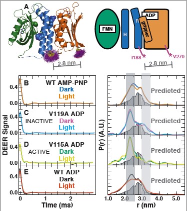
Translation of environmental cues into cellular behavior is a fundamental, necessary process in all forms of life. In bacteria, this process frequently involves two-component systems in which a sensor histidine kinase (HK) autophosphorylates in response to a stimulus before subsequently transferring the phosphoryl group to a response regulator that controls downstream effectors. Many details of the molecular mechanisms of HK activation are still unclear, due to complications associated with the multiple signaling states of these large, multidomain proteins. To address these challenges, we used ESR and complementary solution biophysical approaches to examine the conformational changes upon activation of a minimal, blue-light-sensing histidine kinase from Erythrobacter litoralis HTCC2594, EL346, uncovering new molecular details of the activation of HKs. Our data show that multiple conformations coexist in the dark state of EL346 in solution, which may explain the enzyme's residual dark state activity. We also observed that activation involves destabilization of the helices in the dimerization and histidine phosphotransfer-like (DHpL) domain, where the phosphoacceptor histidine resides, and their interactions with the catalytic domain. Similar light-induced changes occur to some extent even in constitutively active or inactive mutants, showing that light sensing can be decoupled from activation of kinase activity. These structural changes mirror those inferred by comparing X-ray crystal structures of inactive and active HK fragments, suggesting that they are at the core of conformational changes leading to HK activation. More broadly, our findings uncovered surprising complexity in this simple system and allow us to outline a mechanism of the multiple steps of HK activation. Funding: R01TM106239 (KHG); P41GM103521 (JHF); F32GM119311 and Burroughs Wellcome Fund (ID). Publication: Proc. Natl. Acad. Sci. U.S.A 116, 4963-4972 (2019); PMC6421462. |
|
|
|
|
Igor Dikiy (Structural Biology Initiative, CUNY Advanced Science Research Center, New York, NY) Uthama Edupuganti (Structural Biology Initiative, CUNY Advanced Science Research Center, New York, NY, Biochemistry Ph.D. Program, Graduate Center, CUNY, New York, NY) Rinat Abzalimov (Structural Biology Initiative, CUNY Advanced Science Research Center, New York, NY) Peter P. Borbat (ACERT) Madhur Srivastava (ACERT, Meinig School of Biomedical Engineering, Cornell University, Ithaca, NY) Jack H. Freed (ACERT) Kevin H. Gardner (Structural Biology Initiative, CUNY Advanced Science Research Center, New York, NY, Biochemistry, Chemistry and Biology Ph.D. Programs, Graduate Center, CUNY, New York, NY, Department of Chemistry and Biochemistry, CCNY, New York, NY) |
|
|
|
About ACERT Contact Us |
Research |
Outreach |
ACERT is supported by grant 1R24GM146107 from the National Institute of General Medical Sciences (NIGMS), part of the National Institutes of Health. |
|||||
| ||||||||