.svg) National Institute of General Medical Sciences |
 |
 |
National Biomedical Resource for |
| ACERT's Service and Collaborative Projects | ||
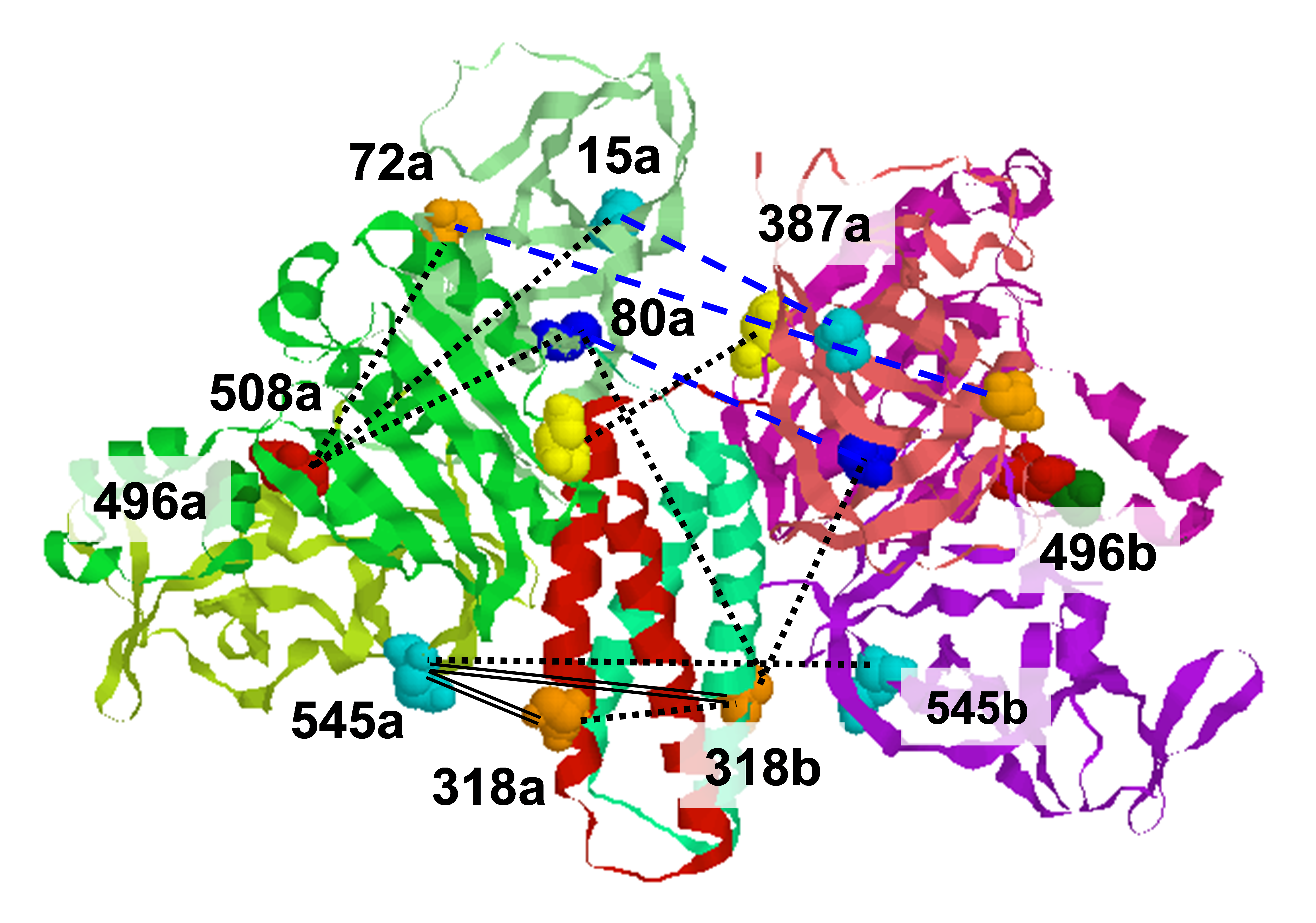
Ebola virus disease is a serious global health concern given its periodic occurrence, high lethality, and the lack of approved therapeutics. Certain drugs that alter intracellular calcium, particularly in endolysosomes, have been shown to inhibit Ebola virus infection; however, the underlying mechanism is unknown. We provided evidence that Zaire ebolavirus (EBOV) infection is promoted in the presence of calcium as a result of the direct interaction of calcium with the EBOV fusion peptide (FP). We identified the glycoprotein residues D522 and E540 in the FP as functionally critical to EBOV's interaction with calcium. We showed using ESR and biophysical assays that interactions of the fusion peptide with Ca2+ ions lead to lipid ordering in the host membrane during membrane fusion, and these changes are promoted at low pH and can be correlated with infectivity. This study shows that calcium directly targets the Ebola virus fusion peptide and influences its conformation. As these residues are highly conserved across the Filoviridae, calcium's impact on fusion, and subsequently infectivity, is a key interaction that can be leveraged for developing strategies to defend against Ebola infection. Funding: NSF 1504846 (to SD and GRW); P41GM103521 and R01GM123779 (to JHF); Samuel C. Fleming Family Graduate Fellowship and NSF Graduate Research Fellowship (to LN). Molecular graphics generated using the Chimera package, P41GM103311 to the Resource for Biocomputing, Visualization, and Informatics, UCSF. Publication: ACS Infect. Dis. 6, 250-260 (2020); PMC7040957. |
||
|
||
|
Lakshmi Nathan (Robert Frederick Smith School of Chemical and Biomolecular Engineering, Cornell University, 120 Olin Hall, Ithaca, New York) Alex Liqi Lai (Baker Laboratory, Department of Chemistry and Chemical Biology, Cornell University, Ithaca, New York) Jean Kaoru Millet, Marco R. Straus (Department of Microbiology and Immunology, College of Veterinary Medicine, Cornell University, Ithaca, New York) Jack H. Freed (Baker Laboratory, Department of Chemistry and Chemical Biology, Cornell University, Ithaca, New York) Gary R. Whittaker (Department of Microbiology and Immunology, College of Veterinary Medicine, Cornell University, Ithaca, New York) Susan Daniel. (Robert Frederick Smith School of Chemical and Biomolecular Engineering, Cornell University, 120 Olin Hall, Ithaca, New York) |
||
|
|
About ACERT Contact Us |
Research |
Outreach |
ACERT is supported by grant 1R24GM146107 from the National Institute of General Medical Sciences (NIGMS), part of the National Institutes of Health. |
|||||
| ||||||||