.svg) National Institute of General Medical Sciences |
 |
|
National Biomedical Resource for |
| ACERT's Service and Collaborative Projects | |
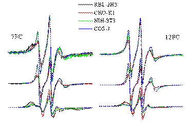
A number of studies have indicated that eukaryotic cell membranes contain coexisting liquid-ordered and liquid-disordered lipid domains. However, the current evidence for such phase separation is indirect, and there had been no direct demonstration of differences in the ordering and dynamics for the lipids in these two types of regions or their relative population in the plasma membranes of live cells. By means of ESR studies employing several chain-labeled phosphatidylcholine spin labels we have succeeded in providing direct evidence for the presence of two different types of lipid populations in the plasma membranes of whole, live RBL-2H3 mast cells, Chinese hamster ovary (CHO-K1) cells, COS-7 cells, and mouse NIH-3T3 cells. Analysis of the ESR spectra recorded from 5°C to 37°C, show that the spin-labeled lipids experience two different types of environments, a liquid-ordered(Lo)-like and a liquid-disordered(Ld)-like with distinct order parameters, and rotational diffusion rates in the two environments, and with some differences amongst the four cell types. The spectra of 7PC and 12PC in the plasma membranes of these cells at 25°C are shown in the Figure (top panel). The Ld-like and Lo-like components obtained from the analysis, are shown in the middle and bottom panels, respectively. These results suggest that coexistence of lipid domains that differ significantly in their dynamic order in the plasma membrane is a general phenomenon. The (Lo)-like region, is typically found to be the major component, which is in contrast to the “raft” model that has been proposed for the plasma membrane, wherein small Lo lipid rafts exist in a “sea” of disordered lipids, but it is consistent with a model of a continuous Lo-like phase in the outer leaflet of the plasma membrane. It is likely that the Ld lipid population could be due to the presence of transmembrane proteins, which disturb the order in the lipids in their surrounding environment and make them more mobile. The existence of distinct ESR spectra from the two environments are consistent with the sizes of the different regions being no smaller than about 20 Å, but we do not rule out small-scale variations of the order of a few lipid molecules in size in the Lo-regions, whose effects could be averaged out on the ESR time-scales. Publication: M.J. Swamy, L. Ciani, M. Ge, A.K. Smith, D. Holowka, B. Baird, and J.H. Freed, Biophys. J., 90, 4452-4465 (2006); PMC1471862 |
|
|
|
|
M. J. Swamy, (Dept. of Chemistry, University of Hyderabad) L. Ciani, (Dept. of Chemistry, University of Florence) D. Holowka, B. Baird (Dept. of Chemistry and Chemical Biology, Cornell University), M. Ge, J. H. Freed (ACERT) |
|
|
|
About ACERT Contact Us |
Research |
Outreach |
ACERT is supported by grant 1R24GM146107 from the National Institute of General Medical Sciences (NIGMS), part of the National Institutes of Health. |
|||||
| ||||||||