.svg) National Institute of General Medical Sciences |
 |
|
National Biomedical Resource for |
| ACERT's Service and Collaborative Projects | |
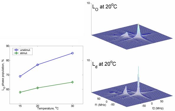
We report one of the first demonstrations of changes in lipid membrane structure and dynamics upon response to external stimuli in a 2D-ELDOR study of plasma membrane vesicles (PMV) from RBL cells that are stimulated by antigen crosslinking of the IgE receptors. We have shown that 2D electron-electron double resonance (2D-ELDOR) is significantly more sensitive to the dynamics than is cw-ESR through the homogeneous broadening and the evolution of the cross-peaks as a function of mixing time. Thus it is naturally sensitive to the changes in the phase structure in biomembranes. Experiments were performed on the PMV isolated from RBL-2H3 mast cells with and without antigen crosslinking of IgE receptors at 15, 20, and 30°C. Small but significant changes are observed in the 2D-ELDOR spectra upon stimulation, which could not be detected by cw ESR. To achieve this observation, we performed spectral fitting in the full Sc- domain (i.e. in the real and imaginary parts of the Fourier-transformed signals instead of in the magnitude format). We were thus able to recover the absorption mode spectrum which provides the best resolution to subtle spectral changes. Our results show that the spectra are best fit with two spectral components (see below), indicating coexistence of liquid-ordered (Lo) and liquid-disordered (Ld) phases. The phase structure changes due to stimulation and its temperature variation were carefully examined according to the dynamic parameters reported from the 16-PC spin label. We find that the population of the Lo phase is decreased upon stimulation, indicating the lipid environment is remodeled to be more disordered (see below). In addition the motional rate, R| and the order parameter, S0 decrease slightly for both components. This is consistent with a model wherein the IgE receptors, which aggregate upon stimulation, lead to more lipids in the Ld phase that are associated with the receptor aggregation, and their dynamic and structural properties are altered. Publication: Y.-W. Chiang, A. Costa-Filho, B. Baird, and J.H. Freed, J. Phys. Chem. B, 115, 10462-10469 (2011); PMC3165081 |
|
|
|
|
Yun-Wei Chiang, Jack H. Freed, (ACERT) Barbara A. Baird, David Holowka (Cornell Univ.), Antonio J. Costa-Filho (Univ. de São Paulo, Brazil.) |
|
|
|
About ACERT Contact Us |
Research |
Outreach |
ACERT is supported by grant 1R24GM146107 from the National Institute of General Medical Sciences (NIGMS), part of the National Institutes of Health. |
|||||
| ||||||||