.svg) National Institute of General Medical Sciences |
 |
|
National Biomedical Resource for |
| ACERT's Service and Collaborative Projects | |
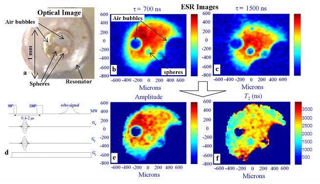
ESR microscopy is an emerging technique, aimed at obtaining spatially-resolved information with micron resolution of samples containing paramagnetic species. The potential applications of this technique include medical tissue examinations and the biophysics of live cells. One of our first practical applications is, in the field of slow chemical (e.g. drug) release. Recently, there has been extensive use of polymeric microspheres as a matrix for the slow release of drugs inside the body. To model and subsequently control this process, one requires tools for observing the drug distribution and monitoring the physical conditions within the sphere, after preparation, and during the release process. Currently the major tool used for such measurements is laser scanning confocal fluorescence microscopy, which employs fluorescent labeled drugs. The problems with this method are that it does not enable one to penetrate deep into the sphere, it provides a non-linear image intensity (due to unknown absorption and scattering coefficients in the sphere), and it cannot be employed easily during the in-vitro/in-vivo release process. Furthermore fluorescence does not have a good capability to quantify the porosity of the spheres, and the self diffusion tensor of the molecules in the sphere. ESR microscopy however, which is a new magnetic resonance imaging method developed in our lab, has the potential of overcoming the limitations of optical methods. We have examined several types of polymer microspheres with a typical size of 100 microns, internalized with stable organic radicals. We successfully monitored the 3D radical distribution during the release process and measured the spatially resolved T2 of the radicals with a typical resolution of ~10 microns. The figure below shows an optical image of the spheres soaked in trityl radical solution (a) and imaging results (b-c) measured with the pulse sequence (d) with typical microwave pulse separations, τ, of 700 and 1500 ns. These images enables us to obtain amplitude (e) and T2 images (f), which show not only a predictable reduction in signal inside the sphere (due to the polymer matrix volume), but also significant reduction in T2 inside the sphere, which is attributed to an increased viscosity (possibly due to the presence of poly-ethylene-glycol inside the sphere). Here only the central sphere absorbed water efficiently. Further investigations along these lines would help to explain the kinetics of the release process, and may aid in the development of better methods of sphere preparation for more controlled release. Publications: |
|
|
|
|
Aharon Blank, Curt Dunnam, Peter Borbat, Jack H. Freed (ACERT) Naraharisetti Pavan Kumar, and Chi-Hwa Wang (Natl. Univ. of Singapore) |
|
|
|
About ACERT Contact Us |
Research |
Outreach |
ACERT is supported by grant 1R24GM146107 from the National Institute of General Medical Sciences (NIGMS), part of the National Institutes of Health. |
|||||
| ||||||||