.svg) National Institute of General Medical Sciences |
 |
 |
National Biomedical Resource for |
| ACERT's Service and Collaborative Projects | ||
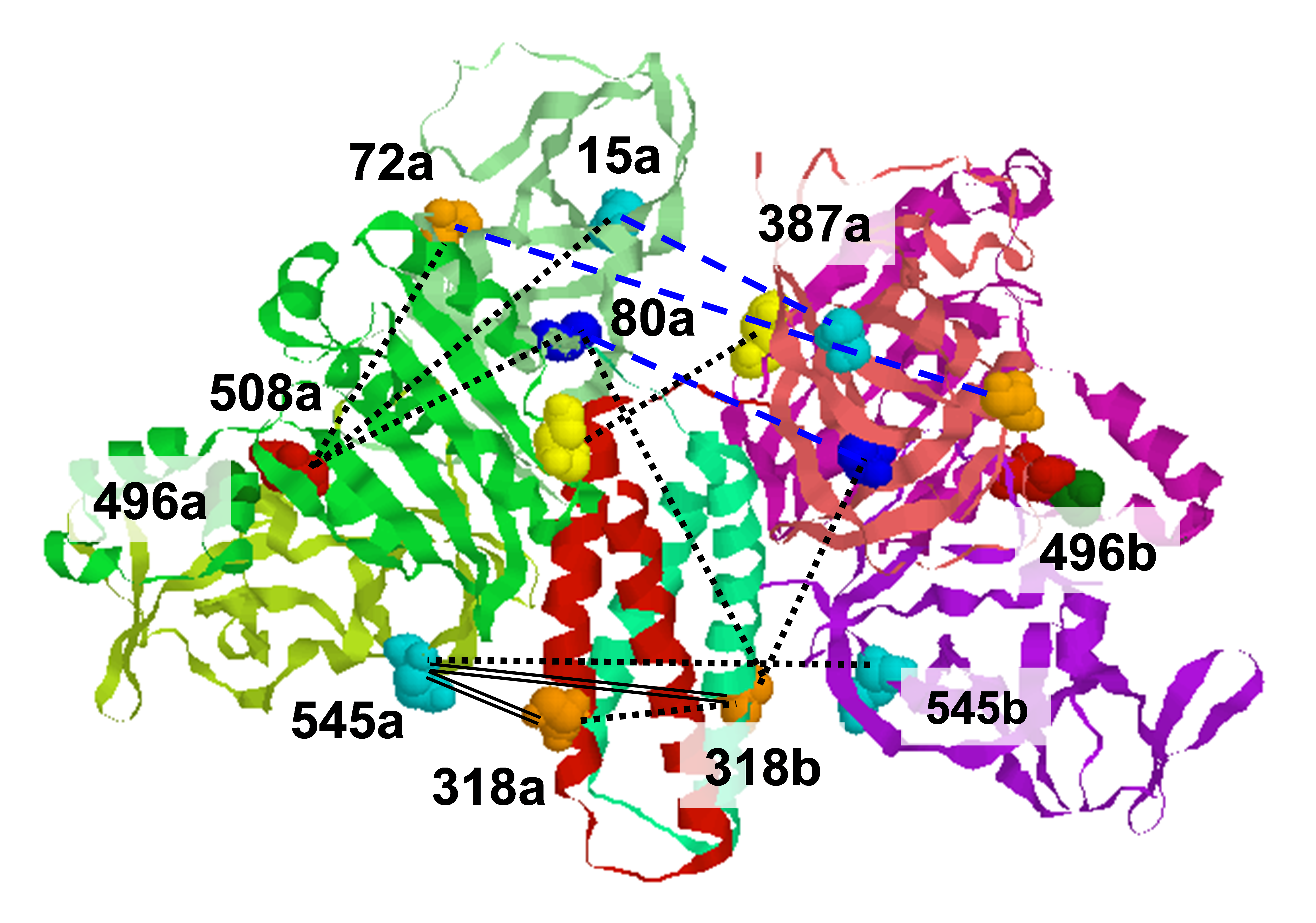
The protein alpha-synuclein (αS) is linked to both sporadic and familial Parkinson's disease (PD) through its appearance in Lewy bodies (intraneuronal deposits that constitute a diagnostic hallmark of PD) and through several genetic polymorphisms (three point mutations and gene triplication or duplication) that lead to early onset disease. αS functions. Detergent micelles induce a similar structural transition in αS, and the sodium dodecyl sulfate (SDS) micelle-bound form of the protein has been used as a surrogate for the vesicle-bound state in a number of structural studies employing solution state NMR. When bound to SDS micelles, the membrane-interacting N-terminal domain of αS adopts two segments of helical structure separated by an ordered linker, and they are oriented anti-parallel to one another but do not contact each other (see illustration below). We use pulsed dipolar ESR spectroscopy to measure directly inter-helix distances in both SDS and lyso-1-palmitoylphosphotidylglycerol (LPPG) micelles. The four-pulse DEER signal is shown below for the 3/83 mutant for SDS (dashed) and LPPG (solid), and the distance distributions, P(r) obtained by Tikhonov regularization is also shown below. We obtained a matrix of distances (see below) that characterizes the SDS micelle-bound state of the protein. When SDS is replaced with LPPG we found that the two αS helices splay further apart from each other. All inter-helix distances confirm an anti-parallel orientation of the two helices in both micelle types. The structural differences observed in LPPG-bound αS may be a consequence either of a change in the local conformation of the linker region, which may be sensitive to the surrounding headgroups, or of a difference in the size and/or geometry of the micelle formed by the longer LPPG acyl chains. The latter should lead to a micelle with a larger radius or major/minor semi-axes. Thus, an appealing explanation for our data is that the topology of micelle-bound αS is determined by that of the bound micelle. Publication: P.P. Borbat, T.F. Ramlall, J.H. Freed, and D. Eliezer, J. Am. Chem. Soc., 128, 10004-05 (2006); no PMCID |
||
|
||
|
P. P. Borbat, J. H. Freed (ACERT) Trudy F. Ramlall, and David Eliezer (Weill Medical College) |
||
|
|
About ACERT Contact Us |
Research |
Outreach |
ACERT is supported by grant 1R24GM146107 from the National Institute of General Medical Sciences (NIGMS), part of the National Institutes of Health. |
|||||
| ||||||||