.svg) National Institute of General Medical Sciences |
 |
|
National Biomedical Resource for |
| ACERT's Service and Collaborative Projects | |||
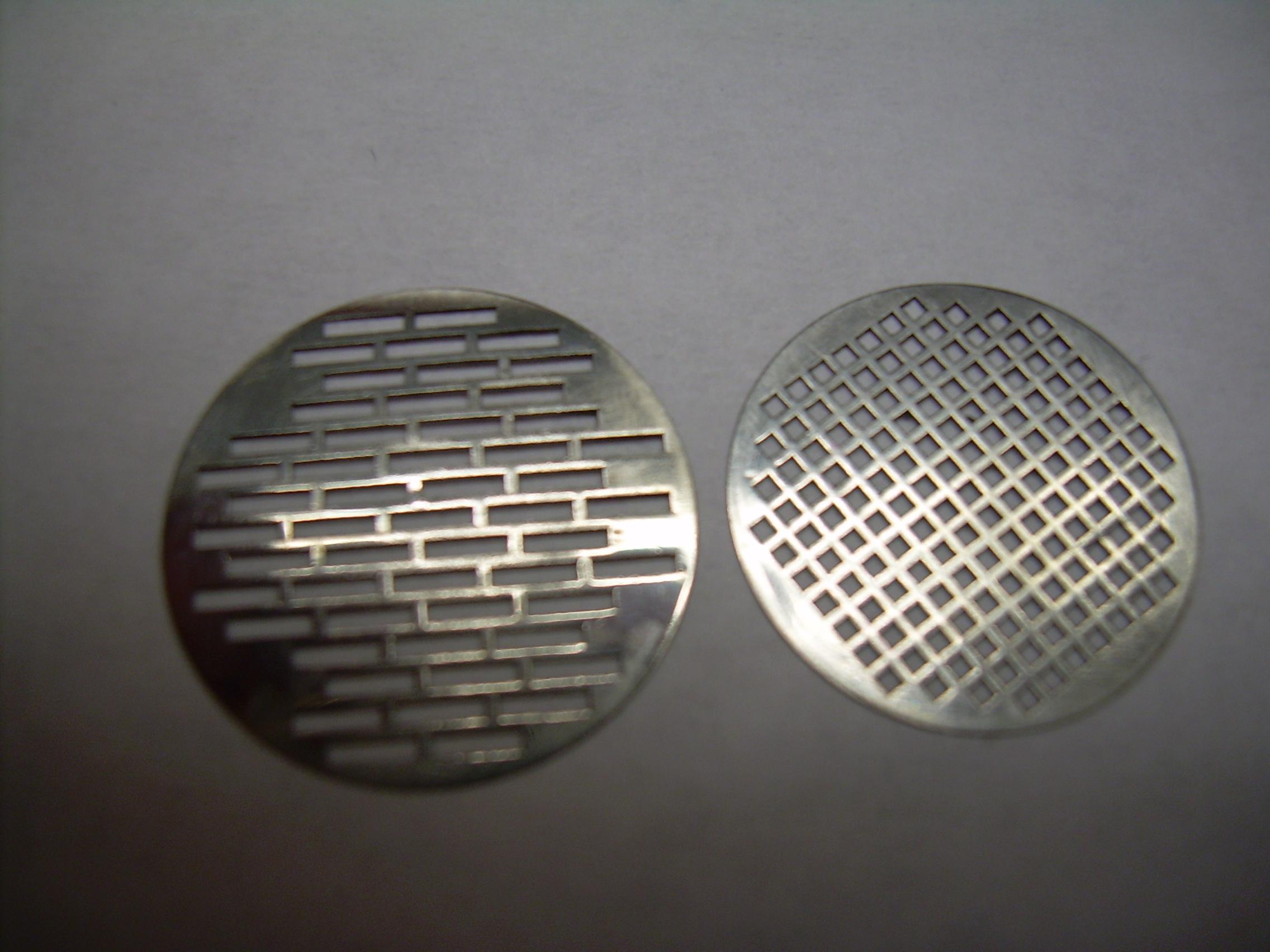
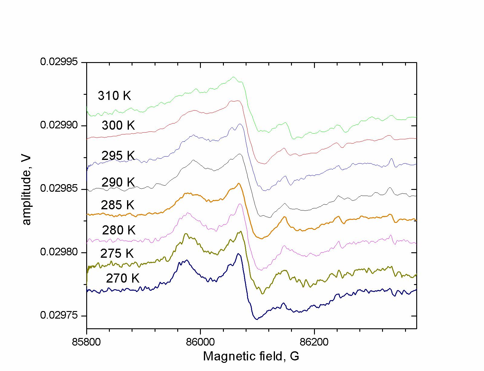
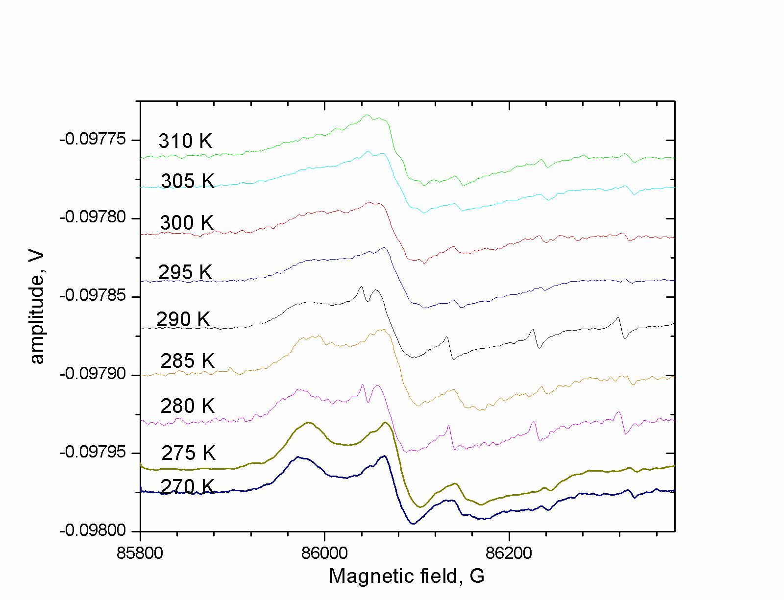
Studies of spin-labeled proteins in aqueous solution under physiological conditions offer the opportunity to obtain important insights into protein dynamics. These insights can subsequently inform models of protein function. In order to make full use of the information available in these spectra, it is important to have excellent concentration sensitivity, as high concentrations of solution proteins, e.g. > 100 microMolar, have a tendency to aggregate, except under exceptional circumstances. At ACERT we have successfully addressed this challenge by improving the coupling of the Fabry-Perot resonator and by developing a method to increase the volume of aqueous samples contained in the FP resonator. The coupling is determined by the reflectivity of the mirror at the input to the resonator. To obtain partial reflectivity, we construct this mirror with a metallic mesh of appropriate dimension. Instead of constructing the mesh with a series of small square apertures of specified size and spacing, we have introduced the novel idea of rectangular meshes, shown in the figure below left. Since the incident radiation is linearly polarized, rotation of such a rectangular mesh changes the degree of reflectivity of the beam, making it possible to critically couple the resonator. Critical coupling of the resonator maximizes the signal. This enables us to detect microMolar levels of motionally narrowed nitroxide spectra with just a single (30 sec) spectral scan at 240 GHz. Increasing the amount of sample in the high magnetic field (and low electric field) regions of the resonator can also increase the concentration sensitivity. The best shape of a lossy aqueous sample is a thin disk of solution placed perpendicular to the mm. wave beam in a position of minimum electric field. We have found that two such samples placed in adjacent minima of the E-field in the FP resonator leads to a doubling of the signal intensity and signal-to-noise. These new design features were successfully applied to a study of HIV-protease in buffered aqueous solution. The concentration of the protein was no more than 100 microMolar. We have compared the spectra at 240 GHz in the presence, and in the absence, of inhibitor, as shown below in the center and right respectively. The inhibitor is a drug that interferes with the protein-cleaving action of the protease, and thus viral propagation. The improved sensitivity allowed us to obtain spectra that show a clear difference between the two cases, unlike the spectra at 9 GHz, which are identical. It is anticipated that careful analysis of these results will lead to a better understanding of the protein dynamic changes resulting from the influence of the drug. Publication: D.S. Tipikin, K.A. Earle, and J.H. Freed, Appl. Magn. Reson., 37, 819-832 (2010); PMC2865681 |
|||
|
|||
|
D.S. Tipikin, K.A. Earle, J.H. Freed (ACERT) L. Galiano, G. Fanucci (Department of Chemistry, University of Florida, Gainesville, FL) |
|||
|
|
About ACERT Contact Us |
Research |
Outreach |
ACERT is supported by grant 1R24GM146107 from the National Institute of General Medical Sciences (NIGMS), part of the National Institutes of Health. |
|||||
| ||||||||