.svg) National Institute of General Medical Sciences |
 |
|
National Biomedical Resource for |
| ACERT's Service and Collaborative Projects | ||
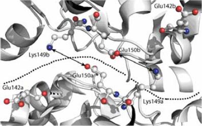
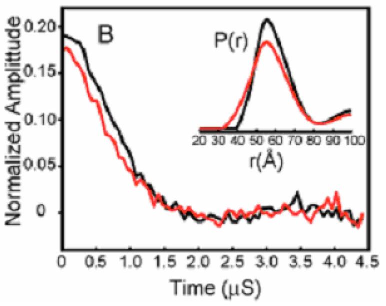
Monoamine oxidases (MAO’s) are outer mitochondrial membrane (OMM)-bound flavoenzymes. Their functions include the oxidative deamination of neurotransmitters including serotonin, adrenaline and noradrenalin (MAOA), dopamine (both MAOA and MAOB), phenylethylamine and other trace amines (MAOB). These oxidation processes are important in maintaining physiological levels of monoamine neurotransmitters in neuronal cells and dietary amines in other tissues. Age-related increase of MAOA and MAOB activities have been implicated in several neurological disorders including Parkinson’s disease and Alzheimer’s disease (MAOB), and chronic depression (MAOA). Selective MAO inhibitors are therefore clinically proven drug targets for treating these disorders. MAOA has been of particular interest in recent studies because of its involvement in cognitive development in humans. Deletion of the MAOA gene results in phenotypes of mental retardation and aggressive behavior in humans and in mice. The 3D-crystal structures of human and rat MAOA (rMAOA), and hMAOB show similar protein folds, however, different oligomeric forms. That is, hMAOA crystallizes as a monomer, whereas both hMAOB and rMAOA are dimeric in their crystalline forms. Based on the sequence analyses of human and other mammalian MAOA’s (including non-human primates) and modeling studies, it has been proposed that hMAOB and rMAOA exist in the dimeric form due to the presence of a conserved Glu-X residue, (with X being 142 in MAOB’s, or 151 in MAOA’s), which facilitates the formation of two Glu-Lys salt bridges at their dimer interface (see figure below left). These salt bridges are destabilized in hMAOA due to a human exclusive Glu151Lys mutation, which causes unfavorable Lys-Lys electrostatic repulsion, and thus promotes the monomeric form of hMAOA in the crystal. Since both MAOA and MAOB are outer mitochondrial membrane (MOM)-bound proteins, their properties are best studied in the intact mitochondria or in the MOM-bound forms. We applied pulsed dipolar ESR spectroscopy (PDS) to elucidate the oligomeric states of human and rat MAO’s and their Glu-X to Lys or Lys-X to Glu mutants in the MOM. We used a synthetic MAO-specific spin-labeled inhibitor, named ParSL which covalently binds to the flavin cofactor at the active site of MAO, forming a N5-flavocyanine adduct. The figure below shows the PDS data on ParSL-labeled wild-type hMAOA (black line), and rMAOA (red line). The dipolar signals obtained from the ParSL-labeled hMAOA sample appear nearly identical to those obtained for rMAOA, and hMAOA Lys151Glu mutant. All samples show an average inter-spin distance of 56±2Å. These distances are in good agreement with that estimated from the clorgyline-inhibited rMAOA crystal structure, indicating that hMAOA, rMAOA, and hMAOA Lys151Glu mutants are all dimeric in the MOM. These data demonstrate that the X-ray crystallographic structure of hMAOA does not reflect its oligomeric state in the MOM. This is the first PDS study on a membrane protein in its natural membrane environment, and it demonstrates the utility of this novel technique for double spin labeling for determining the state of oligomerization and probing protein structures. Publication: A.K. Upadhyay, P.P. Borbat, J. Wang, J.H. Freed, and D.E. Edmondson, Biochemistry, 47, 1554-1566 (2008); no PMCID |
||
|
||
|
A. K. Upadhaya (Departments of Biochemistry and Chemistry, Emory University School of Medicine, Atlanta) P.P Borbat, J. Wang, J.H. Freed, D. E. Edmondson (ACERT) |
||
|
|
About ACERT Contact Us |
Research |
Outreach |
ACERT is supported by grant 1R24GM146107 from the National Institute of General Medical Sciences (NIGMS), part of the National Institutes of Health. |
|||||
| ||||||||