.svg) National Institute of General Medical Sciences |
 |
|
National Biomedical Resource for |
| ACERT's Service and Collaborative Projects | |||
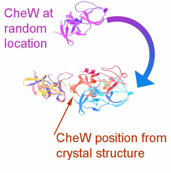
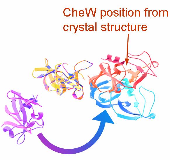
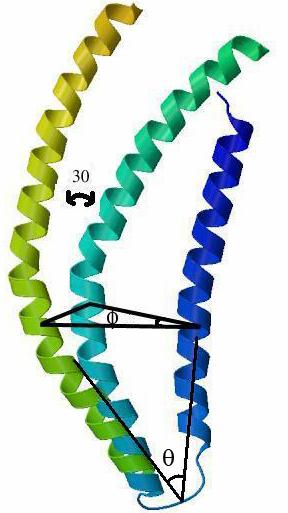
The modeling of protein–protein complexes greatly benefits from the incorporation of experimental distance restraints. Pulsed dipolar electron spin resonance spectroscopy is one such powerful technique for obtaining long-range distance restraints in protein complexes. Measurements of the dipolar interaction between two spins placed specifically within a protein complex give information about the spin–spin separation distance. We have developed a convenient method to incorporate such long-range distance information in the modeling of protein–protein complexes that is based on rigid body refinement of the protein components with the software Crystallography and NMR System (CNS). The use of 4 to 5 different labeling sites on each protein component provides sufficient coverage for producing accuracies limited by the uncertainty in the spin-label conformation within the complex. With an asymmetric scheme of allocating this uncertainty, we showed the importance of longer distances within a limited set of total restraints. This method has been applied to the refinement of the complex formed between the histidine kinase, CheA and its coupling protein, CheW. It is illustrated in the figure at the left below. Starting with random orientations of the two proteins, the program gives a final conformation close to what is present in the crystal structure of the P5/CheW complex. The method has also been applied to the refinement of intra-helical separations in the protein a-synuclein bound to micelles, which is illustrated in the figure at the right below. This figure provides a comparison of helix orientations in the a-synuclein from the ESR-refinement and NMR. The orientation of the two anti-parallel a-synuclein helixes (residues 3-34 and 44-94) derived from the NMR is shown in blue. Superposition of the N terminal of helix from the rigid body refined structure, places the second helix rotated by angle of 30° with respect to the NMR structure. Publication: J. Bhatnagar, J.H. Freed, B.R. Crane, Methods Enzymol., 423, 117-133 (2007); no PMCID |
|||
|
|||
| J. Bhatnagar, J.H. Freed, and B. R. Crane (ACERT) | |||
|
|
About ACERT Contact Us |
Research |
Outreach |
ACERT is supported by grant 1R24GM146107 from the National Institute of General Medical Sciences (NIGMS), part of the National Institutes of Health. |
|||||
| ||||||||