.svg) National Institute of General Medical Sciences |
 |
|
National Biomedical Resource for |
| ACERT's Service and Collaborative Projects | ||||||||||||||||||||||||||||||||||||||||||||||||||||||||||||||||||||||||||||||||||||||||||||||||||||||||||
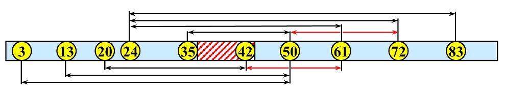
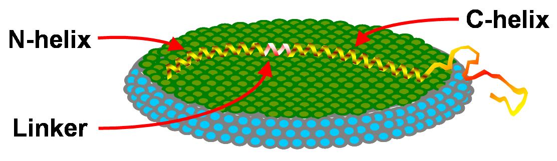
Alpha-synuclein (αS) is a highly conserved presynaptic protein that participates in synaptic strength maintenance and dopamine homeostasis. It is also involved in regulation of intracellular dopamine levels at several points of control. However, accumulation of αS amyloid fibrils was implicated as the major reason in the development of Parkinson's disease (PD). It was shown by NMR that in detergent micelles the protein adopts two extended surface-bound helices separated by a non-helical linker, that the helices are oriented in an antiparallel fashion, and that no inter-helical contacts are formed. In previous pulsed dipolar ESR (PDS) distance measurements of αS bound to different sized micelles we showed that the helices splay further apart on the surface of larger micelles. We have now used PDS to measure distances in αS bound to lipid vesicles, rod-like micelles, and isotropic lipid bicelles, all of which present the protein with a more extensive, less highly curved surface than spheroidal micelles. To circumvent problems associated with vesicles and micelles, we developed the use of PDS with lipid bicelles, which provide a true lipid-bilayer structure, yet have a particle size nearly as small as that of micelles and insure high lipid concentration and bilayer surface area. Our primary objective was to obtain long distance constraints. Distances measured for αS between labels placed from 15 to 59 residues (Figure) apart are shown in the Table. They are in close agreement with those expected for a single continuous helix. In fact, the average distance per residue (cf. Table) is within ±1% of that for an α-helix, which argues strongly for a single, unbroken helix, as depicted in the Figure below. Our results suggest that when αS is bound to synaptic vesicles (its natural binding target in vivo), it is the extended helix conformation of αS that predominates. Publication: E.R. Georgieva, T.F. Ramlall, P.P. Borbat, J.H. Freed, and D. Eliezer, J. Am. Chem. Soc., 130, 12856-12857 (2008); PMC2626176 |
||||||||||||||||||||||||||||||||||||||||||||||||||||||||||||||||||||||||||||||||||||||||||||||||||||||||||
|
||||||||||||||||||||||||||||||||||||||||||||||||||||||||||||||||||||||||||||||||||||||||||||||||||||||||||
|
Elka R. Georgieva, (Department of Chemistry and Chemical Biology, Cornell University, Ithaca, NY 14853) Trudy F. Ramlall, (Department of Biochemistry and Program in Structural Biology, Weill Cornell Medical College, 1300 York Avenue, New York, NY 10065) Peter P. Borbat, Jack H. Freed (Department of Chemistry and Chemical Biology, Cornell University, Ithaca, NY 14853) David Eliezer (Department of Biochemistry and Program in Structural Biology, Weill Cornell Medical College, 1300 York Avenue, New York, NY 10065) |
||||||||||||||||||||||||||||||||||||||||||||||||||||||||||||||||||||||||||||||||||||||||||||||||||||||||||
|
||||||||||||||||||||||||||||||||||||||||||||||||||||||||||||||||||||||||||||||||||||||||||||||||||||||||||
|
About ACERT Contact Us |
Research |
Outreach |
ACERT is supported by grant 1R24GM146107 from the National Institute of General Medical Sciences (NIGMS), part of the National Institutes of Health. |
|||||
| ||||||||