.svg) National Institute of General Medical Sciences |
 |
|
National Biomedical Resource for |
| ACERT's Service and Collaborative Projects | |
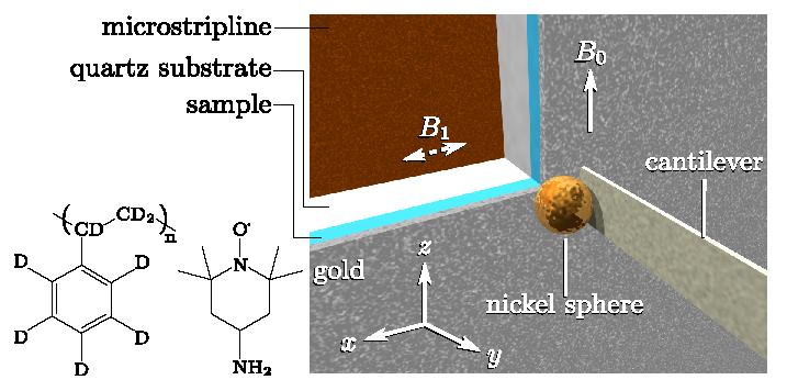
We have developed an approach that extends the applicability of ultrasensitive force gradient detection of magnetic resonance to samples with spin-lattice relaxation times (T1) as short as a single cantilever period. To demonstrate the generality of the approach, which relies on detecting either cantilever frequency or phase, we used it to detect electron spin resonance from a T1 = 1 ms nitroxide spin probe (cf. Figure) in a thin film at 4.2 K and 0.6 T. Using a custom fabricated cantilever with a 4 μm diameter nickel tip, we achieve a magnetic resonance sensitivity of 410 Bohr magnetons in a 1 Hz bandwidth. Our theory for this quantitatively predicts both the lineshape and the magnitude of the observed cantilever frequency shift as a function of field and cantilever-sample separation. Good agreement was found between nitroxide T1's measured mechanically and inductively, indicating that the cantilever magnet is not an appreciable source of spin-lattice relaxation here. We suggest that the new approach has a number of advantages that make it well suited to push magnetic resonance detection and imaging of nitroxide spin labels in an individual macromolecule to single-spin sensitivity. In the figure below, we show the schematic of the scanned-probe electron spin resonance experiment. A microstripline half-wave resonator delivers a transverse magnetic field, B1, oscillating at 17.7 GHz. In the center of the resonator, the microwave field oscillates along the x direction. A longitudinal Zeeman field of magnitude B0 ~ 0:6 T is applied along the z axis. The high-compliance cantilever has its long axis along y and oscillates in the x direction. The cantilever's 4 μm-diameter nickel tip was affixed by hand. The sample is a 230 nm-thick film of 40 mM TEMPAMINE in perdeuterated polystyrene, coated with 20 nm of gold. The sample film was spin-coated onto a 250 μm-thick quartz wafer. For clarity, sample and substrate are not drawn to scale. Publication: E.W. Moore, S. Lee, S.A. Hickman, S.J. Wright, L.E. Harrell, P.P. Borbat, J.H. Freed, and J.A. Marohn, Proc. Natl. Acad. Sci. U.S.A., 106, 22251-22256 (2009); PMC2799694 |
|
 |
|
|
Eric W. Moore, Sang Gap Lee, Steven A. Hickman, Sarah J. Wright, (Dept. of Chemistry and Chemical Biology, Cornell University, Ithaca, NY 14853) Lee E. Harrell, (Dept. of Physics, U.S. Military Academy, West Point, NY 10996) Peter P. Borbat, Jack H. Freed, and John A. Marohn (Dept. of Chemistry and Chemical Biology, Cornell University, Ithaca, NY 14853) |
|
|
|
About ACERT Contact Us |
Research |
Outreach |
ACERT is supported by grant 1R24GM146107 from the National Institute of General Medical Sciences (NIGMS), part of the National Institutes of Health. |
|||||
| ||||||||
