.svg) National Institute of General Medical Sciences |
 |
|
National Biomedical Resource for |
| ACERT's Service and Collaborative Projects | |||||||
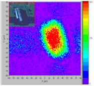
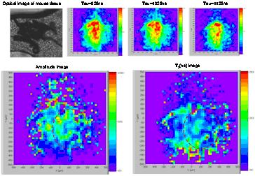
High resolution electron spin resonance microscopy (ESRM) can provide a 3-Dimensional view of medically relevant information such as oxygen concentration, viscosity, and diffusion coefficients in biological systems. In magnetic resonance imaging, resolution is typically limited by the number of available spins in a single imaging voxel and molecular diffusion. However, due to the shorter time scale, ESRM resolution is less affected by the diffusion. The ESRM instrument developed at ACERT can detect as small as ~107 spins (NMR microscopy can detect ~1012 spins), and can produce very intense gradient pulses, permitting sub-micron resolution. A high spin sample, i.e. LiPc crystal, which has ~108 electron spins per μm3, can be considered as an ideal sample for demonstrating this resolution. An ESR image with sub-micron resolution was obtained as shown in Figure 1(a), with resolution of ~0.7x0.75x7.5 μm or a single imaging voxel size of 4 μm3. This is an improvement of ~20 over our earlier published results. The peak gradient strength is now 75 T/m for X and 47 T/m for Y. For biomedical applications with water soluble spin probes, such as Trityl, the available spin concentration in 1mM solution is ~6x10⁵ spins per μm3. Thus, the number of spins available in a single voxel limits the resolution obtainable in reasonable imaging times. As an example mouse leg tissue with PC3 tumor was imaged as shown in Figure 1(b). The estimated resolution is ~20x20x20 μm (we achieve even better resolution in some other cases). After taking ESRM images with different τ's, an amplitude image and T2 image can be generated as shown in the bottom row. After calibration with a known sample, the oxygen concentration image in the tissue can be generated from the T2 image. These images can also be used to calibrate standard EPR imaging, which may not show the heterogeneity of the Trityl concentrations in the tissue, due to its lower resolution (ca. 1mm). Publication: C.S. Shin, C.R. Dunnam, P.P. Borbat, B. Dzikovski, E.D. Barth, H.J. Halpern, and J.H. Freed, Nanosci. Nanotechnol. Lett., 3, 561-567 (2011); PMC3188420 |
|||||||
|
|||||||
|
Chang S. Shin, Curt R. Dunnam, Peter P. Borbat, Boris Dzikovski (National Biomedical Center for Advanced ESR Technology, Dept of Chemistry and Chemical Biology, Cornell University) Gene Barth, Howard Halpern (Department of Radiation and Cellular Oncology, University of Chicago) Michael Shklyar (Schulich Faculty of Chemistry, Technion–Israel Institute of Technology) Jack H. Freed (National Biomedical Center for Advanced ESR Technology, Dept of Chemistry and Chemical Biology, Cornell University) |
|||||||
|
|||||||
|
About ACERT Contact Us |
Research |
Outreach |
ACERT is supported by grant 1R24GM146107 from the National Institute of General Medical Sciences (NIGMS), part of the National Institutes of Health. |
|||||
| ||||||||