.svg) National Institute of General Medical Sciences |
 |
|
National Biomedical Resource for |
| ACERT's Service and Collaborative Projects | |
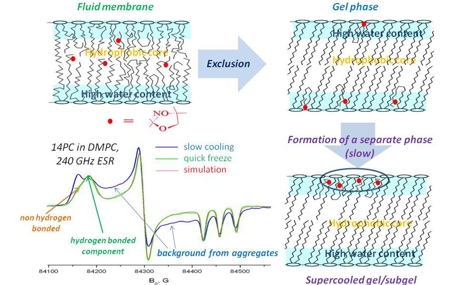
Lipid spin labels containing nitroxide groups at different positions in the fatty acid chain, such as n-PC spin labels, are a useful and proven tool in lipid research. Through a vast number of studies, they have provided crucial insights into the structure of model and biological membranes, reported on the membrane fluidity, polarity, phase state and presence of microscopic domains, accessibility of different depth positions in the lipid bilayer for oxygen and other polar and non-polar paramagnetic compounds. In this project we revisited the use of PC spin labels for studying polarity gradients in model and biological lipid bilayers. With superior g-factor resolution of our 240 GHz ESR we were able to resolve spectral components unresolved at lower frequencies and to answer questions about using PC spin labels in membrane studies which were unanswered for the last two decades. We have shown that the ESR parameters of PC spin labels in frozen membranes do not simply represent the membrane polarity or water penetration profile. Instead, they show a distribution between hydrogen-bonded (HB) and non-hydrogen bonded (non-HB) states, which is affected by a number of factors in the membrane composition. Similar to the exclusion of solutes from crystallizing solvents, the pure bulk gel phase excludes nitroxides, forcing acyl chains to take bent conformations. In these conformations the nitroxide is hydrogen-bonded. Furthermore, upon gradual cooling in the supercooled gel, PC labels undergo slow lateral aggregation resulting in a broad background signal. However, if the sample is instantly frozen, this background is replaced by the HB component. In membranes with cholesterol the observed HB/non-HB ratio can best be described by a partition-like equilibrium between nitroxides located in defects of lipid structure within the hydrophobic core and those close to the membrane surface. This is illustrated in the Figure. Publication: B. Dzikovski, D. Tipikin, and J.H. Freed, J. Phys. Chem. B, 116, 6694-6706 (2012); PMC3376253. |
|
|
|
|
Boris Dzikovski (ACERT) Dmitriy Tipikin (ACERT, Department of Community and Family Medicine, Dartmouth Medical School, Hanover, NH) Jack H. Freed (ACERT) |
|
|
|
About ACERT Contact Us |
Research |
Outreach |
ACERT is supported by grant 1R24GM146107 from the National Institute of General Medical Sciences (NIGMS), part of the National Institutes of Health. |
|||||
| ||||||||