.svg) National Institute of General Medical Sciences |
 |
|
National Biomedical Resource for |
| ACERT's Service and Collaborative Projects | ||
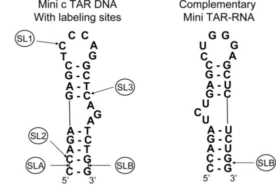
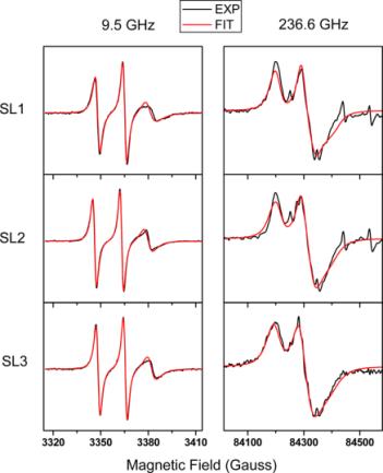
This study was directed to understand the internal dynamics of a model stem.loop oligonucleotide from HIV-1 and the change in these dynamics upon its interaction with HIV-1 nucleocapsid protein NCp7. The stem.loop structure is found in the TAR (transactivation response) region of c TAR DNA and TAR RNA. As shown both in vivo and in vitro, the binding of NCp7 inhibits self-priming within such a stem.loop structure and promotes annealing for the formation of duplexes between complementary TAR RNA and TAR DNA. EPR at 236.6 and 9.5 GHz probed the tumbling of nitroxide spin probes in the lower stem, in the upper loop, and near the bulge of mini c TAR DNA. High-frequency 236.6 GHz EPR, not previously applied to spin-labeled oligonucleotides, was notably sensitive to fast, anisotropic, hindered local rotational motion of the spin probe, occurring approximately about the NO nitroxide axis. The spin-label studies with 240 GHz EPR provided information about rapid subnanosecond, hindered, local anisotropic motions and showed differences in these motions. The most rapid motion was in the lower stem. High-frequency EPR showed a dynamic difference between the end-labeled mini c TAR DNA and the mini TAR RNA and the ψ3 RNA, implying a more ordered environment of the label at 5 °C in the two RNAs. These dynamic differences among stem, loop, and bulge and between mini c TAR DNA and mini TAR RNA provide insight into oligonucleotide dynamics, and they point to future comparative RNA.DNA studies. The slowing of nanosecond tumbling motions of large segments of the oligonucletide, which are best observed by 9.5 GHz EPR, was enhanced by increasing the nucleocapsid protein NCp7:mini c TAR DNA ratio from zero to one to two. A greater slowing was observed from the labels in the loop and near the bulge of mini c TAR DNA. This differential dynamic sensitivity to the binding of NCp7, as probed by EPR, is thus a functional aspect of mini c TAR. Publication: Y. Sun, Z. Zhang, V.M. Grigoryants, W.K. Myers, F. Liu, K.A. Earle, J.H. Freed, and C.P. Scholes, Biochemistry, 51, 8530-8541 (2012); PMC3549007. |
||
|
||
|
Yan Sun (Department of Chemistry, University at Albany) Ziwei Zhang (ACERT, State Key Laboratory of Magnetic Resonance and Atomic and Molecular Physics, Wuhan Institute of Physics and Mathematics) Vladimir M. Grigoryants (Department of Chemistry, University at Albany) William K. Myers (Department of Chemistry, University at Albany, and Department of Chemistry, University of California--Davis) Fei Liu (Department of Chemistry, University at Albany) Keith A. Earle (Department of Physics, University at Albany) Jack H. Freed (ACERT) and Charles P. Scholes (Department of Chemistry, University at Albany) |
||
|
|
About ACERT Contact Us |
Research |
Outreach |
ACERT is supported by grant 1R24GM146107 from the National Institute of General Medical Sciences (NIGMS), part of the National Institutes of Health. |
|||||
| ||||||||