.svg) National Institute of General Medical Sciences |
 |
|
National Biomedical Resource for |
| ACERT's Service and Collaborative Projects | ||
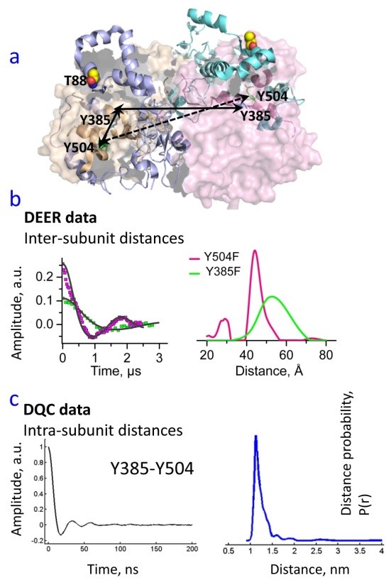
Cyclooxygenase-2 (COX-2) is a dimeric monotopic membrane protein that catalyzes the first step in prostaglandin synthesis by converting arachidonic acid into prostaglandin H2. Each monomer contains physically distinct cyclooxygenase and peroxidase active sites, which are functionally linked via a heme moiety. Non-steroidal anti-inflammatory drugs and non-substrate fatty acids can bind to one monomer of the dimer and allosterically regulate catalysis in the partner monomer. Previous biochemical studies have demonstrated that the enzyme functions with half-of-sites reactivity such that at any time only one monomer of the dimer is catalytically active, yet multiple tyrosyl (Tyr) radicals are involved in COX catalysis. However, their roles in one-sided reactivity and allosteric regulation have not been established. We completed a study based on several pulse dipolar ESR measurements aimed to determine the partitioning of Tyr radicals between the protomers of the COX-2 dimer. We applied double electron-electron resonance (DEER) and double-quantum coherence ESR (DQC) to measure the distances between Tyr radicals produced in the COX-2 catalytic cycle. Our studies revealed that the radicals are formed from both Tyr385 and Tyr504 residues and in each half. This was determined by measuring Tyr-Tyr radical distances in wild type protein or COX-2 constructs in which either Tyr385 or Tyr504 were mutated out for phenylalanine. Thus, we learned that the half-sided reactivity of COX-2 dimer is not a result of catalytically-active tyrosine radical being generated in only one of the monomers. Furthermore, Tyr504 radical, which is non-catalytic, is generated in both monomers of the dimer, thereby supporting an early proposed idea that this radical may serve as a "radical reservoir" replenishing oxidizing equivalents to the catalytic Y385 after depletion with reductants. Our findings contribute significantly to the understanding of the COX-2 functional mechanism. This study required us to measure very short (11 Å) and very long (up to 65 Å) distances between Tyr, which are positioned very close (~10 - 15 Å) to fast-relaxing iron ions. Publication: |
||
|
||
|
Benjamin J. Orlando and Michael G. Malkowski (Hauptman-Woodward Medical Research Institute and Department of Structural Biology, State University of New York, Buffalo) Elka R. Georgieva, Peter P. Borbat, Jack H. Freed (ACERT) |
||
|
|
About ACERT Contact Us |
Research |
Outreach |
ACERT is supported by grant 1R24GM146107 from the National Institute of General Medical Sciences (NIGMS), part of the National Institutes of Health. |
|||||
| ||||||||