.svg) National Institute of General Medical Sciences |
 |
|
National Biomedical Resource for |
| ACERT's Service and Collaborative Projects | ||||||
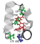
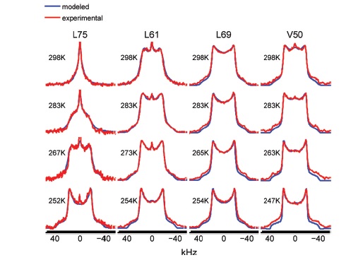
Deuterium lineshape analysis of CD3 groups has emerged as a particularly useful tool for studying μs - ms protein motions in the solid-state. The models devised so far consist of several independently conceived simple jump-type motions. They are comprised of physical quantities encoded in their simplest form; improvements are only possible by adding yet another simple motion, thereby changing the model. The various treatments developed are case-specific; hence comparison amongst the different systems is not possible. We have developed a new methodology for 2H NMR lineshape analysis free of these limitations. It is based on the microscopic-order-macroscopic-disorder (MOMD) approach. In MOMD motions are described by diffusion tensors, spatial restrictions by potentials/ordering tensors, and geometric features by relative tensor orientations. Jump-type motions are recovered in the limit of large orientational potentials. Model-improvement is accomplished by monitoring the magnitude, symmetry and orientation of the various tensors. The generality of MOMD makes possible comparison amongst different scenarios. CD3 lineshapes from the Chicken Villin Headpiece Subdomain, and the Streptomyces Subtilisin Inhibitor, are used as experimental examples. All of these spectra are reproduced by using rhombic local potentials constrained for simplicity to be given by the L = 2 spherical harmonics, and axial diffusion tensors. Potential strength and rhombicity are found to be ca. 2 - 3 [kBT]. The diffusion tensor is tilted at 120o from the C-CD3 axis. The perpendicular (parallel) correlation times for local motion are 0.1 - 1.0 ms (3.3 - 30 μs). Activation energies in the 1.1 - 8.0 kcal/mol range are estimated. Future prospects include extension to the 2H relaxation limit, application to the 15N and 13C NMR nuclei, and accounting for collective motions and anisotropic media. |
||||||
|
||||||
|
Eva Meirovitch (Mina and Everard Goodman Faculty of Life Sciences, Bar-Ilan University, Israel) Zhichun Liang and Jack H. Freed (ACERT) |
||||||
|
|
About ACERT Contact Us |
Research |
Outreach |
ACERT is supported by grant 1R24GM146107 from the National Institute of General Medical Sciences (NIGMS), part of the National Institutes of Health. |
|||||
| ||||||||