.svg) National Institute of General Medical Sciences |
 |
|
National Biomedical Resource for |
| Multifrequency Studies |
|
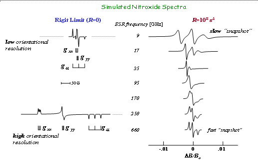
Multifrequency ESR is a powerful tool for studying the structures and dynamics of biological systems. By measuring ESR spectra at different frequencies, various dynamics of different correlation times can be selectively probed. That is, higher the ESR frequency, faster the motional dynamics is “captured” in the ESR spectrum: for a given rotational diffusion rate the spin label motion appears to become slower as one utilizes higher frequencies. For the same rotational rate, at low or conventional frequencies (e.g. 9 GHz) one may observe motionally narrowed spectra, whereas at high frequencies (e.g. 250 GHz) the spectra may display very slow motion features, almost the rigid limit character. This "snapshot" feature enables a multi-frequency ESR approach to study the complex modes of motion of proteins, DNA, and other polymers, which leads to decomposition of the motion into modes according to their different time scales.
|
|
|
|
|
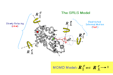
Moreover, as the ESR frequency increases, there is the increase of the orientational resolution of the nitroxide spectrum, due to the dominant role of the g-tensor. For rigid limit spectra at 250 GHz, one can clearly distinguish the well-separated spectral regions corresponding to those nitroxide spin labels with their x-axes parallel to B0, their y-axes parallel to B0, and their z-axes parallel to B0. Then as motion is introduced (e.g. by warming the sample) one can discern the axis (or axes) about which the motion occurs. Because of this enhanced resolution, the 250 GHz slow-motional spectra are much more sensitive to the details of the motional dynamics than are those at microwave frequencies. This dynamics-orientation information can be untangled from spectra with the aid of our models, “Slowly Relaxing Local Structure” (SRLS) and "Microscopic Order - Macroscopic Disorder” (MOMD) , provided one chooses the right frequencies and designs the experiment wisely. A good example is our work with T4 Lysozyme. |
|
|
|
|
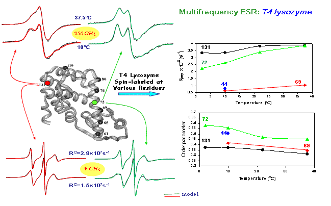
We used high-frequency (e.g. 250 GHz) to "freeze-out" overall tumbling motions, (and other slow motions). This provided us with dramatic sensitivity to the faster "local” motions of tether and the attached to it spin label: local ordering, local diffusion tensor, geometry. This allowed us to see even subtle differences in motions of the spin label attached at different sites of T4 lysozyme. 9 GHz spectra, it turn provided us with the better sensitivity to the overall tumbling rate of the T4 lysozyme
|
|
|
|
|
molecule. In particular, see figure, we studied T4 lysozyme labeled at sites 72 and 131. These sites are of particular interest, since the nitroxide side-chain makes no tertiary contacts, i.e. no interactions with nearest neighbor side-chains. This results in quite similar local motional rates at both sites. However, their 250 GHz spectra are qualitatively different. One should note that site 72 is at the center of the long helix, while site 131 is in a short two-turn helix. One thus expects site 72 to have a higher ordered helix surface than site 131. This results in higher local ordering at site 72 than site 131 and, in consequence, the observed difference in 250 GHz spectra. The spectra from all labeled sites can be fit to the same overall tumbling rate, Rc. |
|
© 2022 |
|
About ACERT Contact Us |
Research |
Outreach |
ACERT is supported by grant 1R24GM146107 from the National Institute of General Medical Sciences (NIGMS), part of the National Institutes of Health. |
|||||
| ||||||||