Friday, Feb. 16, 2024
2pm-7pm
701 Clark Hall |
|
Saturday, Feb. 17, 2024
9am-2pm
401 Physical Sciences Building (PSB) |
| 1:00 |
Registration and Coffee |
|
9:00 |
Registration, Coffee, and Welcome |
| 1:15 |
Welcome & History of ESR (Dr. Madhur Srivastava) |
|
|
|
| 1:30 |
Introduction to ESR: How ESR Works
Prof. Jack Freed |
|
|
|
| 2:00 |
Applications of ESR
Prof. Brian Crane |
|
|
|
| 2:30 |
Session 1 |
(20 min talk, 5 min Q&A) |
|
9:20 |
Session 3 |
(20 min talk, 5 min Q&A) |
| 2:30 |
A. Pulse Dipolar ESR: Its Many Applications to Biomolecular Structure and Function I |
|
9:20 |
A. How Viruses Initially Attack Host Cells (e.g., SARS-2) |
| 2:55 |
B. Detecting and Identifying Transient Radicals: Chemical Intermediates and Biochemical Processes |
|
9:45 |
B. Watching Microsecond Chemical Exchange Processes in Real Time |
| 3:20 |
C. Multifrequency ESR: Variable Time Shots of Protein Dynamics and More |
|
10:10 |
C. Pulse ESR Methods to Study the Dynamic Structure of Proteins and their Membrane Interactions |
| 3:45 |
D. Blood-based Biomarkers for Disease Detection: ALS (Amyotrophic Lateral Sclerosis) Case Study |
|
10:35 |
D. Forensics, Dosimetry, Archeology, and More: Ubiquitous ESR |
| 4:10 |
Break |
|
|
11:00 |
Break |
|
|
| 4:20 |
Session 2 |
(20 min talk, 5 min Q&A) |
|
11:20 |
Demo and Tour Session Rotations |
(30 min each) |
|
| 4:20 |
E. How To Study Intrinsically Disordered Proteins |
|
|
Continuous Wave ESR |
(Alex Lai and Timotheé Chauviré) |
Baker B27 |
| 4:45 |
F. Distance Measurements in Heterogeneous Oligomers |
|
|
Pulsed ESR |
(Rob Dunleavy and Tufa Assafa) |
Baker B20 |
| 5:10 |
G. In Vivo Studies of Protein Structure and Dynamics in Cells |
|
|
Sample Preparation |
(Boris Dzikovski and Jess Whittemore) |
Baker B19 |
| 5:35 |
H. Plant Science Studies by ESR |
|
|
Tour of ACERT Facilities |
(Madhur Srivastava and Curt Dunnam) |
Baker B6 |
| |
|
|
|
Tour of ACERT Facilities |
(Peter Borbat and Aritro Sinha Roy) |
Baker B31 |
| 6:00 |
Dinner |
700 Clark Hall |
|
1:30 |
Lunch and Talk |
401 PSB |
| |
|
|
|
|
A Practical Approach to Biomolecular Pulse ESR (Thomas Schmidt, Ph.D. – NIH Staff Scientist) |
Friday Sessions
Session A: Pulse Dipolar ESR: Its Many Applications to Biomolecular Structure and Function I–Peter Borbat and Brian Crane
The talk includes a brief introduction to the history of Pulse Dipolar ESR (PDS), its main implementations, and its analogies to NMR methods. A glimpse into numerous PDS applications to biomolecular structure and function highlights its major approaches using multiple examples from works of ACERT and other labs. The instrumentation and spin labeling methods and their limits will also be discussed.
Session B: Detecting and Identifying Transient Radicals: Chemical Intermediates and Biochemical Processes–Alex Lai
A wide range of chemical and biochemical processes produce radicals, such as food deterioration, aging of materials, stress response in cells, and chemical reactions. The degree of those process can be indicated by the amounts of radicals produced, which is measured using Quantitative ESR. The species of radicals can also be identified using ESR, which reveals the mechanism of the reaction. However, as most radicals are unstable and transient, regular room-temperature ESR is not suitable in most cases. Either low temperature ESR or spin trapping ESR is used to “freeze” or “capture” the transient radicals. We will use the direct carboxylation of pyridines using CO2, the thermal degradation of thaumatin (a potent sweet tasting protein), and the stress response against drug in yeast cells as examples.
Session C: Multifrequency ESR: Variable Time Shots of Protein Dynamics and More–Boris Dzikovski
High Field and multifrequency ESR is a useful and powerful tool in biomedical ESR. It provides superior spectral resolution, and often gives a different and complementary views of the system compared to ESR at lower frequencies. In our examples, we show how it is used to study the complex modes of molecular motion of proteins and to resolve different hydrogen bonded states of molecules in the biomembrane environment.
Session D: Blood-based Biomarkers for Disease Detection: ALS (Amyotrophic Lateral Sclerosis) Case Study–Aritro Sinha Roy and Rob Dunleavy
Identifying new disease biomarkers in blood samples by ESR: Experiments & data analysis
ALS is a fast-progressing neurodegenerative disease that cannot be diagnosed at its early stages. We measure the distance distribution between two copper centers in the protein SOD1 by ESR to identify ALS. Challenges associated with detection at blood level concentration of SOD1 have been overcome with our special data analysis technique.
Session E: How To Study Intrinsically Disordered Proteins–Rob Dunleavy and Aritro Sinha Roy
ESR is uniquely suited to characterize both the structure and dynamics of Intrinsically Disordered Proteins. Intrinsically Disordered Proteins (IDPs) and Intrinsically Disordered Regions (IDRs) constitute over 40% of the human proteome. Magnetic resonance spectroscopy (ESR and NMR) is uniquely suited to characterize these challenging cases by providing detailed information on protein structure and dynamics. When combined with site directed spin labeling, ESR reports on the interactions between IDPs and their associated binding partners by revealing dynamical and structural transitions. Here, we cover the applications of ESR to studying IDP/IDR's with specific examples of IDPs involved in human diseases.
Session F: Distance Measurements in Heterogeneous Oligomers–Tufa Assafa and Jess Whittemore
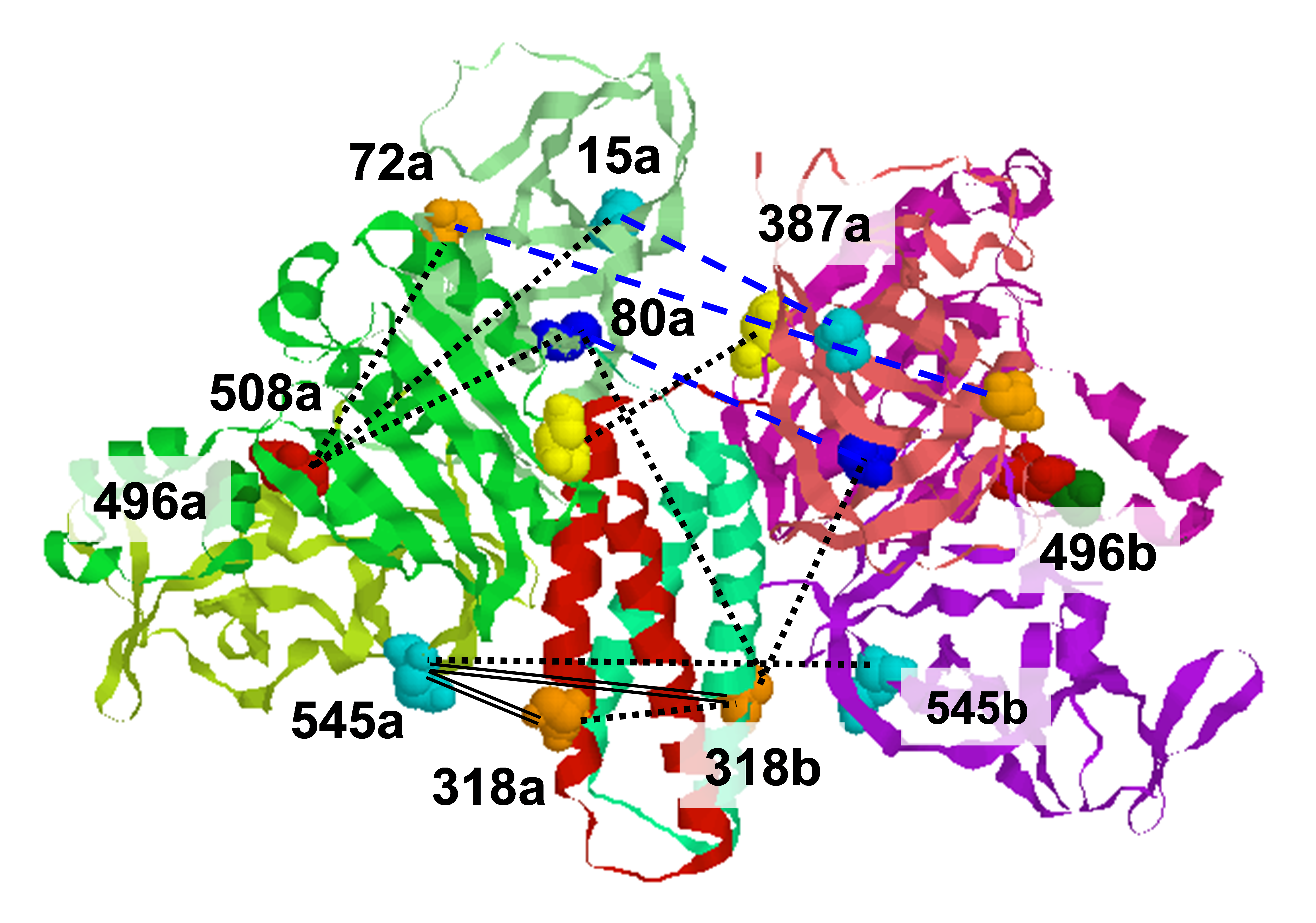
Advances in Pulsed ESR allow the application of distance measurements to larger protein systems, which include hetero- and homo-oligomers. In this session, we will describe the chemical and spectroscopic approaches to extract structural information from these kinds of proteins. We will then show recent applications of these methods to the Aer/CheW/CheA complex.
Session G: In Vivo Studies of Protein Structure and Dynamics in Cells–Timothée Chauviré
In vivo experiments can be challenging but ultimately give more information about the protein/enzyme environment. You will learn what the state of art of in-vivo ESR. We will present the different labelling techniques that have been achieved and their use in in-vivo studies. We will then consider a specific example based on the research at ACERT: ESR characterization of the native flavin spin label of aerotaxis protein in E. Coli cells.
Session H: Plant Science Studies by ESR–Madhur Srivastava
Changes in chemical and biochemical structures can reveal and differentiate between their normal and abnormal function. While diseases in plants can be detected via visual inspection at the later stages, we will show that it is possible to detect early signs of plant disease by using a biochemical approach, where ESR can be used to measure the changes in the activity of paramagnetic species. Examples from vineyards will be used to reveal the biochemical detection, which can be used for other plants.
Saturday Sessions
Session A: How Viruses Initially Attack Host Cells (e.g., SARS-2)–Alex Lai
The COVID-19 pandemic is caused by SARS-CoV-2, which shares the same basic topological structure of a wide variety of viruses, which are a threat to public health such as HIV, influenza, and Ebola. The core of the virus is wrapped by a viral membrane called the viral envelope. Thus, the infection of virus requires a critical step called membrane fusion, which is the fusion between the viral envelope and the host's membranes, and thus releases the viral genetic materials into the host cells. The application of ESR technology elucidates how this process is mediated by the viral Fusion Peptide, which is a critical domain consisting of 20-30 residues and is responsible for the initialization of the membrane fusion. The understanding of the mechanism gives hints for therapy strategies. We will also show how this mechanism is "borrowed" by some host cells during evolution to achieve important physiological functions.
Session B: Watching Microsecond Chemical Exchange Processes in Real Time–Boris Dzikovski
2D ESR is a new tool for studying rapid chemical exchange. While 2D NMR is an established routine method to study chemical exchange, until recently the use of ESR for this purpose was limited. The new 2D ELDOR technique combined with High Field ESR allows for real-time observation of exchange processes on a microsecond to nanosecond time scale which is not accessible by other 2D physical methods.
Session C: Pulse ESR Methods to Study the Dynamic Structure of Proteins and their Membrane Interactions–Peter Borbat and Brian Crane
The focus is on applications of pulse ESR to peptides, proteins, and lipid membranes, local ordering, and dynamics, as well as membrane-protein interactions based on snapshots of immobilized proteins, as well as in real-time dynamic states at physiological temperatures. Particular emphasis will be on 2D-FT methods, such as 2D-ELDOR.
Session D: Forensics, Dosimetry, Archeology, and More: Ubiquitous ESR–Madhur Srivastava
Contamination from impurities is detrimental to human health. However, impurities serve a useful purpose based on the origin of the substance. While the chemical structure of a substance remains the same across different samples, the impurities associated with it vary based on how the sample was produced and its location. ESR techniques can effectively reveal the origins of impurities, by reliably studying the composition and characteristics of the impurities. Several compounds have free radicals as impurities. For example, we can analyze Schedule I substances that include cannabis, heroin, and cocaine.

The workshop for new ACERT users is supported by the National Institute Of General Medical Sciences of the National Institutes of Health under Award Number 1R24GM146107.
|

